


Research and current projects
Photoacoustic Imaging and Spectroscopy
The research activities of the Photoacoustic (PA) Imaging and Spectroscopy Group are focussed on the development of the instrumentation, methodologies, and genetic reporters for molecular an functional imaging through an interdisciplinary research programme that is conducted in collaboration with groups at the Martin-Luther-Universität Halle-Wittenberg and other institutions involving physicists, engineers, life scientists and clinicians.
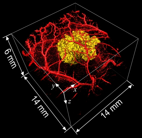
PA image of a melanin expressing tumour and the surrounding normal vasculature.
Overview
- Photoacoustic Imaging and Spectroscopy
- Hybrid forward models for quantitative photoacoustic tomography of blood oxygen saturation
- Highly sensitive biomedical photoacoustic tomography with homodyne optical ultrasound detection
- Photoacoustic lifetime imaging of fluorescent contrast agents using pump-probe excitation
- Functional single-chain polymer nanoparticles for biomedical photoacoustic tomography
- Quantitative photoacoustics for transcranial monitoring of human brain function
- Pulsed laser diodes for photoacoustic imaging of the human brain
- PhotoHEAR - Development of technology for a photoacoustic hearing aid
- Diagnostic Tools and New Treatment Strategies for Individualized Therapy of Colorectal Cancer
- Time resolved fluorescence tomography
- Fluorescent molecules in cancer diagnostics
- GreenSight Smart Monitoring for Sustainable Algal Biotechnology
Hybrid forward models for quantitative photoacoustic tomography of blood oxygen saturation
Biomedical photoacoustic (PA) tomography is based on the generation of ultrasound in the tissue via the absorption of intensity-modulated light. Due to the dominant absorption of haemoglobin in the visible and near-infrared wavelength range, it provides high-resolution 3D images of the vasculature in soft tissue. It is also sensitive to exogenous and genetically expressed contrast agents. A major potential of this technology lies in the ability to recover 3D high resolution images of absolute chromophore concentrations (e.g. oxy- and deoxyhaemoglobin, contrast agents) as well as derived functional parameters, such as blood oxygen saturation. The development of methods for quantitative PA tomography is of crucial importance for the translation of this technology to routine applications in functional and molecular imaging. In this project, comprehensive forward models will be developed to predict multispectral PA images that closely agree with those acquired using different tomographic PA scanner platforms. The models will be used in adjoint form in new inversion algorithms, which will be experimentally validated on images of tissue phantoms acquired using novel experimental methods. The limitations of the new methods will be evaluated in silico. It is envisaged that the models developed in this project will enable true quantitative PA tomography using both model-based and data-driven inversions. The successful development of these models and methods will provide a basis for rapid translation of functional and molecular PA imaging to applications in biology, the life sciences and medicine.
Investigators: Guo Tang (PhD candidate), Jan Laufer (PI)
Funder: Deutsche Forschungsgemeinschaft (DFG) - Project number LA3273/7-1
Highly sensitive biomedical photoacoustic tomography with homodyne optical ultrasound detection
The objective of this project is the development of a purely optical 3D imaging system for biomedical photoacoustic (PA) tomography, characterized by ultra-sensitive acoustic detection, high imaging frame rates, and high spatial resolution. To achieve these goals, existing imaging technologies will be extended through the integration of adaptive optics, enabling maximal noise suppression and, consequently, maximal acoustic sensitivity. These innovations aim to simultaneously optimize key imaging parameters critical for clinical translationnamely imaging depth, frame rate, and contrastwithin a single tomographic system.
Investigators: Jan Sievers (PhD candidate), Jan Laufer (PI), - in cooperation with Claus Villringer (TU Wildau)
Funder: European Regional Development Fund (ERDF)
Photoacoustic lifetime imaging of fluorescent contrast agents using pump-probe excitation
Biomedical photoacoustic (PA) tomography is based on the generation of ultrasound in tissue via the absorption of intensity-modulated light. Due to the dominant absorption of haemoglobin in the visible and near infrared wavelength range, high-resolution 3D images of the vascular networks in soft tissue can be acquired. The PA effect also provides contrast to exogenous contrast agents. Current experimental and computer-aided methods for determining the spatial contrast agent distribution from PA images are limited in their sensitivity, specificity and general validity. In this project, novel experimental and numerical approaches for the detection of fluorophores by pump-probe excitation will be developed and combined with accurate numerical methods to open up a wide range of new applications in multiplexed PA imaging of biophysical and biochemical parameters.
Investigators: Farzin Golmohamadi (PhD candidate), Jan Laufer (PI)
Funder: Deutsche Forschungsgemeinschaft (DFG) - Project number LA3473/8-1
Functional single-chain polymer nanoparticles for biomedical photoacoustic tomography
Molecular photoacoustic (PA) tomography is a hybrid imaging technique that combines strong absorption-based contrast and spectral specificity of purely optical imaging modalities with high spatial resolution of ultrasound. Since the scattering of acoustic waves in tissue is orders of magnitude lower compared to that of light, PA imaging offers high resolution images at depths beyond those offered by purely optical methods. To visualize tissues that are transparent to visible and nearinfrared excitation wavelengths, contrast agents are typically required. The challenge of detection lies in their low abundance, and hence weak contrast, against the overwhelming background of hemoglobin. To enable sensitive detection, new contrast agents are required that possess unique photophysical properties. In addition, novel experimental methods are needed that exploit these properties for detection, thus overcoming the limitations of conventional unmixing approaches. The contrast agents should also offer added functionality, such as the possibility to sense and measure local biophysical and biochemical parameters. In this project, single-chain polymer nanoparticles (SCNP) loaded with dye molecules will be developed for PA and optical imaging. They offer strong absorption, small size, suitable surface properties for targeting, and biocompatibility. Their unique photophysical properties, such as a strong nonlinear PA response, pave the way for the development of highly sensitive and unambiguous detection methods that are based on simple experimental approaches, such as pump-probe excitation. SCNP will also offer a versatile platform for the development of novel contrast agents that act as biosensors of specific chemical species, such as ions or enzymes, or biophysical parameters, such as pH.
Investigators: Marzieh Ezzatpour (PhD), Jan Laufer and Wolfgang Binder (PI)
Funder: Deutsche Forschungsgemeinschaft (DFG) - Project number LA3473/11-1
Quantitative photoacoustics for transcranial monitoring of human brain function
The primary goal of this project is the development of a non-invasive photoacoustic (PA) monitoring method for blood oxygen saturation in the human brain through the intact skull. Photoacoustics is a technique increasingly used in preclinical research as well as some early clinical trials. Photoacoustics uses short light pulses to excite optical absorbers such as blood to emit sound waves. These sound waves can then be measured with ultrasonic sensors. Photoacoustics enables high-resolution measurements of optical absorption at a depth of several centimeters and is primarily used as an imaging modality. Transcranial PA imaging in humans has so far proved challenging to implement. However, transcranial Photoacoustics as a technique not for imaging but local monitoring has, so far, barely been studied. In the proposed project, we would like to investigate and develop instrumentation and algorithms to investigate the feasibility of transcranial PA monitoring of blood oxygen saturation. We are especially interested in the accurate quantitative measurement of physiological blood oxygen saturation in the human brain and will develop a prototype for real-time transcranial PA monitoring for potential functional studies on the human brain.
Investigators: Thomas Kirchner (PI)
Funder: Deutsche Forschungsgemeinschaft (DFG) - Project number 471755457
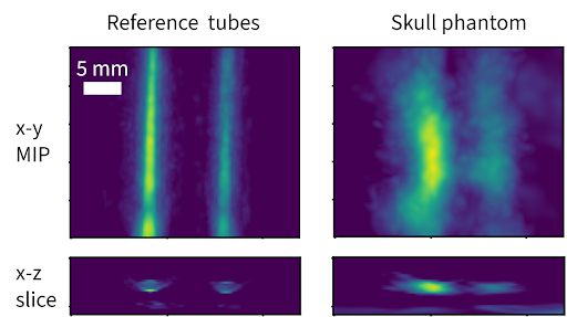
Figure: Artificial blood vessels imaged through thick human skull.
Pulsed laser diodes for photoacoustic imaging of the human brain
Photoacoustic imaging of the human brain has great potential to contribute to rapid diagnosis of strokes and other brain injuries, thus contributing to more effective treatment. Photoacoustics is based on the optical excitation of an ultrasound wave in an optical absorber such as blood. Sudden absorption of nanosecond-long laser pulses generates sound waves in blood vessels - these ultrasound waves are measured and reconstructed on the surface of the tissue by ultrasound sensors, enabling three-dimensional imaging of perfusion. This project focuses on high power laser diode arrays as photoacoustic excitation sources and investigates how these pulsed laser diodes can be used to overcome the technical challenges in transcranial photoacoustic imaging.
Investigators: Thomas Kirchner (PI)
Funder: European Regional Development Fund (ERDF)
PhotoHEAR - Development of technology for a photoacoustic hearing aid
The photoacoustic effect can be used to generate audible sound. This project aims to investigate the feasibility of a photoacoustic-excitation-based bone conduction hearing aid as an alternative or supplement to conventional devices. Bone conduction hearing devices are clinically effective for patients with a functioning inner ear, suffering from conductive or mixed hearing loss. In this work, a modulated laser beam is used to excite sound in the skull, which is conducted to the cochlea in bone. Different excitation and modulation techniques are investigated and evaluated with the goal of attaining sound across the entire audible frequency range for music and speech recognition. To evaluate clinical feasibility, experiments will be performed on a skull phantom that mimics the properties of the human skull.
Investigators: Rajalakshmi Sivarajan (PhD student), Jan Laufer (Co-Investigator), Thomas Kirchner (Co-Investigator) - in collaboration: Dawid Brüning (PhD student), Torsten Rahne (PI)
Funder: European Regional Development Fund (ERDF)
Diagnostic Tools and New Treatment Strategies for Individualized Therapy of Colorectal Cancer
Thera4Age is an interdisciplinary research consortium based at Martin Luther University Halle-Wittenberg (MLU) and focused on developing innovative therapies for age-related diseases. The consortium comprises ten esteemed Principal Investigators and is structured within five sub-projects, each jointly directed by experts from two collaborating disciplines.
Our group is leading the fourth subject in collaboration with Markus Petermanns lab. It involves the comparative evaluation of preclinical models (2D cell cultures, 3D organ-on-a-chip tissue cultures, in vivo models, and computational biology) alongside photoacoustic in vivo imaging. These tools will enable the identification and evaluation of pharmacological targets (including the Serpin family) for colorectal cancer therapy. We are then putting all our photoacoustic imaging expertise to get precise neovascularisation mapping on tumors on different models to monitor the drug efficiency. In addition, we are using pump-probe contrast and fluorescent proteins encoded in reporter genes to further investigate the tumor growth non-invasively.
Investigators: Clement Linger (PostDoc), Jan Laufer (PI) - in collaboration with Markus Petermann
Funder: European Regional Development Fund (EFRE)
Consortium: Thera4Age - Innovative therapy concepts for age-related diseases (Website https://thera4age.uni-halle.de/ / Linkedin https://www.linkedin.com/company/thera4age/ )
Time resolved fluorescence tomography
In this project we use time-resolved fluorescence tomography (FT) for three-dimensional visualization of fluorescent tissues with quantitative depth sensitivity. One goal is the determination of the local pH value by combining time resolved fluorescence with pH sensitive marker molecules. In FT, tissue is excited—typically in the near-infrared window—and the resulting emission is recorded at multiple surface positions and viewing angles. Specific fluorescence markers with pH dependent fluorescence lifetime can be used to generate a 3D pH map of the tissue based on the local fluorescence lifetime (see Figure).
The fluorescence lifetime is widely independent of the chromophore concentration and only marginally distorted by autofluorescent signals so that it represents a much better ruler for the pH as intensity based measurements. Performance is assessed using tissue-mimicking phantoms and ex vivo samples to quantify spatial resolution, sensitivity, contrast-to-noise ratio, and absolute concentration accuracy.
Investigators: Franz-Josef Schmitt (PI), Fabian Rieder (MSc student)
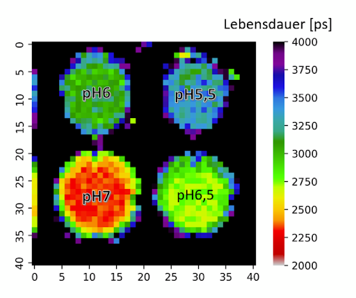
Figure: Lifetime plot of the pH values in a microwell plate
determined with time resolved fluorescence tomography and pH
sensitive marker molecules (fluorescein-isothiocyanate
Fluorescent molecules in cancer diagnostics
We dissect the pH dependence of far-red fluorescent proteins by coupling broadband absorption/emission spectroscopy with multichannel time-correlated single-photon counting (TCSPC) and decay-associated spectra (DAS) analysis. Kinetic models fitted to the DAS reveal the pH dependency of intramolecular relaxation channels and help to understand the interaction of the molecules with protons and support the specific design of optimized pH sensors. Molecular dynamics (MD) at different protonation states rationalize the observed photophysics and give rise to understanding the role of specific amino acids in stabilisation of the chromophore and modulation of the excited-state proton transfer (ESPT) efficiency, providing a mechanistic basis for pH-sensitive lifetime and insensitivity measurements. Collectively, the combined spectroscopy–modeling–MD framework links intramolecular water/chromophore dynamics to ESPT-controlled rates and establishes design rules for red fluorescent protein pH biosensors compatible with lifetime-based readouts, super-resolution/ultrafast fluorescence microscopy, and pump–probe photoacoustic reporting in vivo.
Investigators: Franz-Josef Schmitt (PI)
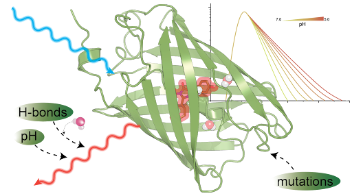
Figure: Targeted mutations enhance fluorescent proteins by
optimizing chromophore stabilisation, chromophore-water-
interaction and excited-state proton transfer for bright
sensors with strong pH sensitivity.
GreenSight Smart Monitoring for Sustainable Algal Biotechnology
GreenSight is a research project focused on harnessing the potential of microalgae to address future global challenges in renewable energy, sustainable food production, and environmental protection. By combining advanced sensor technologies with AI-driven data analysis, GreenSight develops innovative tools to continuously monitor water quality, algal physiology, and growth dynamics in real time.
At the technical level, we are developing distributed sensor systems connected via WiFi. These sensors continuously record environmental parameters such as temperature, CO₂, oxygen, pH, nutrients (NPK), and light absorption. By integrating these data into cloud platforms, we apply machine learning methods to reveal correlations between environmental conditions and algal physiology. This enables us to characterize growth dynamics, identify stress factors, and move toward automated control of cultivation systems.
Our long-term vision is to establish compact, multi-channel optical sensors — such as RGB and hyperspectral chips — as core tools for classifying algal cultures based on their spectral fingerprints. Linking optical signals with physiological models paves the way for real-time metabolic profiling and autonomous, AI-driven photobioreactors. Beyond algal biotechnology, these methods can support environmental monitoring and sustainable agriculture, contributing to the design of intelligent and adaptive biotechnological systems.
Investigators: Franz-Josef Schmitt (PI)
Funder: European Regional Development Fund (ERDF)