Projects
Hybrid forward models for quantitative photoacoustic tomography of blood oxygen saturation (DFG Project Grant LA3273/7-1)
Biomedical photoacoustic (PA) tomography is based on the generation of ultrasound in the tissue via the absorption of intensity-modulated light. Due to the dominant absorption of haemoglobin in the visible and near-infrared wavelength range, it provides high-resolution 3D images of the vasculature in soft tissue. It is also sensitive to exogenous and genetically expressed contrast agents. A major potential of this technology lies in the ability to recover 3D high resolution images of absolute chromophore concentrations (e.g. oxy- and deoxyhaemoglobin, contrast agents) as well as derived functional parameters, such as blood oxygen saturation. The development of methods for quantitative PA tomography is of crucial importance for the translation of this technology to routine applications in functional and molecular imaging. In this project, comprehensive forward models will be developed to predict multispectral PA images that closely agree with those acquired using different tomographic PA scanner platforms. The models will be used in adjoint form in new inversion algorithms, which will be experimentally validated on images of tissue phantoms acquired using novel experimental methods. The limitations of the new methods will be evaluated in silico. It is envisaged that the models developed in this project will enable true quantitative PA tomography using both model-based and data-driven inversions. The successful development of these models and methods will provide a basis for rapid translation of functional and molecular PA imaging to applications in biology, the life sciences and medicine.
Development of highly-sensitive Fabry-Pérot ultrasound sensors
The aim of the project is to increase the acoustic sensitivity of optical ultrasound detectors based on the Fabry-Pérot interferometer and to parallelise this imaging technology. This will enable high frame rate image acquisitions that can be applied to in vivo studies of fast physiological processes. In cooperation with TH Wildau, tunable Fabry-Pérot ultrasound sensors are being developed to enable highly parallelised signal and image acquisition.
Novel genetically expressed reporter proteins and chromophores for photoacoustic imaging of stem cells
Since stem cells are migratory and divide into specialised cell types, genetically expressed reporters are highly advantageous for detection and tracking. While photoacoustic imaging of fluorescent proteins (FP) in small, translucent organisms has been demonstrated, we found many FPs to be unsuitable since their optical absorption coincides with strong haemoglobin absorption. Also, they tend to photobleach, while fluorescence and ground state depopulation reduce the PA signal amplitude.
To overcome these limitations, we pursued the development of methods for the detection of photoswitchable, near-infrared absorbing reporter proteins based on phytochromes. We have recently obtained very encouraging results, which showed that phytochromes can be detected using a dual-wavelength excitation and difference imaging, which completely removes the endogenous contrast. In addition, we found that phytochromes are highly photostable and can be expressed efficiently in mammalian cells.
This allowed deep tissue imaging of growing tumours in a non-invasive and longitudinal manner

Photoacoustic image of the vasculature of a subcutaneous tumor. The tumor cells express a photoswitchable protein (green) and its spatial distribution can be detected by photoacoustic imaging.
Photoacoustic lifetime imaging of fluorescent contrast agents using pump-probe excitation (DFG project grant LA3473/8-1)
Biomedical photoacoustic (PA) tomography is based on the generation of ultrasound in tissue via the absorption of intensity-modulated light. Due to the dominant absorption of haemoglobin in the visible and near infrared wavelength range, high-resolution 3D images of the vascular networks in soft tissue can be acquired. The PA effect also provides contrast to exogenous contrast agents. Current experimental and computer-aided methods for determining the spatial contrast agent distribution from PA images are limited in their sensitivity, specificity and general validity. In this project, novel experimental and numerical approaches for the detection of fluorophores by pump-probe excitation will be developed and combined with accurate numerical methods to open up a wide range of new applications in multiplexed PA imaging of biophysical and biochemical parameters.
Repoter molecules for genetic contrast in molecular photoacoustic tomography- ESF project
The photoacoustic (PA) effect, the conversion of intensity-modulated light into sound, has found application in diverse areas, such as the material sciences, spectroscopy, and deep tissue imaging. It relies on the absorption of optical energy by tissue chromophores, such as haemoglobin, melanin, lipid to generate broadband ultrasound waves that are detected outside the organism and avoids the ionising radiation of x-ray computed tomography to allow longitudinal studies in the same organism without side effects. By exploiting the wavelength-dependence of the optical absorption of oxy- and deoxyhaemoglobin, functional and metabolic imaging has been demonstrated while the use of exogenous and genetically expressed probes has added to molecular imaging capabilities. Genetically expressed proteins and pigment-synthesising enzymes are an attractive approach to produce PA contrasts (e.g. via a photoswitchable phytochromes as shown in Figure 1), and it offers the potential to spy on cells and tumours using PA tomography. To date, comparatively few genetic reporters have been imaged successfully in mammals and their tumour-selective expression in vivo has not been demonstrated.
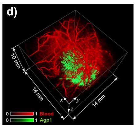
Figure 1. In vivo PA tomography of genetically expressed photoswitchable phytochrome (Agp1) using a Fabry-Pérot-based scanner. 3D image of a subcutaneous tumour expressing a Agp1 (green) and the vasculature (red) of the surrounding tissue.
The objective of th of this ESF-funded project is the development of genetically expressed molecular PA reporters in tumour cells to allow their detection in vivo. It is envisaged that features in the aquired image data sets will allow a correlation of cellular activity with physiological function in deep tissue solid tumours. The Medical Physics research group will design and generate a tumour-specific expression pattern of PA reporters in mammalian cells. Functionalised expression of the PA reporter proteins in malignant tumours will be validated by molecular techniques. Finally, PA tomography is applied to detect tumour-specific PA reporters and produce 3D PA images of tumours in vivo.
The technologies and methods developed in this project will provide novel materials and methods for non-invasive PA reporter gene imaging. It has the potential to enable in vivo studies of the molecular and cellular processes controlling disease progression and to correlate cellular activity with physiological function and anatomical changes. It will have a major impact on a wide range of research fields in the life sciences, biology, and medicine, such as oncology, neurology, and immunology, with applications ranging from basic research to clinical studies.
Project duration: 01.10.2020 - 31.05.2022
Contact: Prof. Dr. Jan Laufer and Dr. Amna Shah Mehmood
Telephone: 0345 55 25400/7
E-mail: jan.laufer@physik.uni-halle.de, amna.mehmood@physik.uni-halle.de